Chapter One: An Acoustics Primer
14. How does the ear work?
This chapter begins our study of psychoacoustics—how the human body and mind perceive the physics of sound discussed previously. We begin with our primary sensorium for sound, the human ear.
 Lars Chittka; Axel Brockmann / CC 2.5
ANATOMY OF THE EAR
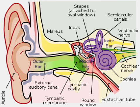
Our primary organ used to sense sound is the ear. We can also hear to a lesser degree through our skull's temporal bones, enabling the function of certain hearing implants along with bone-conduction headphones and smart glasses. The structure of the ear can be divided into three main parts: the outer ear beginning with the pinna (also called the auricle), the middle ear, and the inner ear.
The outer structure of the ear is responsible, in part, for helping us to place the original location of a sound, be it ahead or behind, above or below us. It also helps to funnel and focus sound waves on their way to the middle ear and auditory canal. The outer ear also contains the auditory canal, which terminates in the eardrum, or tympanic membrane. The auditory canal behaves as a closed-tube resonator, amplifying frequencies in the 2-5 kHz range. Attached to the other side of the eardrum, in the middle ear (which is a small space of air), are three tiny bones or ossicles, the malleus, incus and stapes (or hammer, anvil and stirrup). The ossicles connect the ear drum to a fluid-filled inner ear structure called the cochlea at a point called the oval window. The fulcrum/lever design of the ossicles actually amplify the movement transmitted from the ear drum to the oval window of the cochlea by a factor of 15 times or so. If you have ever had a middle ear infection, that air space can fill with fluid, impacting the way you hear. In addition, the Eustachian tube is designed to equalize outside pressure with the middle ear, so when we fly or otherwise rapidly change altitude, until the tube opens to equalize things, our hearing is also impacted.
In the cochlea, the vibrations transmitted from the ossicles to the oval window are then converted into fluid vibrations that move tiny hairs, which are then converted into electrical impulses sent along the auditory nerve (also called the cochlear nerve) to the brain explained in greater detail below. The flexible round window membrane (in the picture above) provides relief for the pressure waves that oscillate through the cochlea's two outer canals. The inner ear, which is surrounded by bone, also contains semicircular canals, which function more for purposes of equilibrium than hearing.
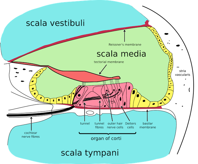 Cochlea Cross-section:
BREAKDOWN OF THE COCHLEA
The cochlea, so named because of its snail-shell-like shape, is a tube that tapers logarithmically (mirroring our perception of pitch, which is also logarithmic), and circles around itself like the scroll of a violin. The scala media in the center divides the tube lengthwise into two sodium-based fluid-filled canals (scala tympani and scala vestibuli), which are joined at the tapered end so that the fluid can reverse direction between them as it reflects from the round window on the other end. The all-important basilar membrane sits on the surface of the potassium-based scala media on the scala tympani side. The ossicles transmit the ear drum's vibration to the cochlea where they attach at the oval window. The resultant pressure waves travel down the fluid of the scala tympani and basilar membrane where they are sensed by the approximately 16-20,000 hair cells called cilia attached to it. These hair cells poke both up and down from a complex structure called the organ of Corti that sits right behind the basilar membrane. The basilar membrane become thinner and less stiff as it approaches the end of the cochlea, which contributes to pitch perception as it transmits its vibrations to the organ of Corti below it. It is the organ of Corti that transforms the stimulated hair cells into nerve impulses. Because of the tapered design of the cochlea, waveforms traveling down the basilar membrane peak in amplitude at differing spots along the way proportional to their frequency, according to the place theory of pitch perception, our current best guess for how we sense higher frequencies.
Higher frequencies peak out at a shorter distance down the tube than lower frequencies. The hair cells at that peak point give us a sense of that particular frequency. It is thought that a single musical pitch is perceived by 10-12 hair cells. Due to the tapered shape of the cochlea, the distance between frequencies follows the same logarithmic distance as our perception of pitch (e.g., the placement of octaves is equidistant). This arrangement is responsible for the fact that a lower frequency at an equal or higher amplitude can mask a higher frequency, but under most circumstances a higher frequency of equal or higher amplitude can't mask a lower frequency. Masking is actually a very complex phenomenon influenced by the pitch and amplitude relationship of the tones inside or outside the critical band, discussed in a later section. The masking phenomenon is used to advantage in reducing the amount of bandwidth and digital memory necessary to produce near-high-fidelity music in compressed perceptually-coded audio formats such as mp3's. Frequencies that would normally be masked are eliminated from the coding altogether, since theoretically they would not be heard or missed.

FACTOID #6: Noise-induced Hearing Loss (NIHL)
Hearing loss caused by exposure to sound is referred to as noise-induced hearing loss. It can result from occasional or even one-time exposure to extremely loud sounds. It can also result from long-term exposure to moderately loud sound, for example from occupational background noise. It is believed long-term exposure to sound above 85 dBA can cause NIHL. While instant damage can occur at 140 dB, pain or the lack of it is not a good indicator of the danger to human hearing. Excellent noise level meters are available for smart phones and often indicate short and longterm exposure safety. The primary mechanism for loss is permanent damage to the cilia and hair cells of the cochlea, which die and do not regenerate. In addition, overexposure causes inflammation in the cochlea that results in poor blood flow to these structures. The loss may manifest itself as a notch in the frequency spectrum, with widening and deepening as the loss becomes more profound. The best strategies for avoiding hearing loss is avoidance of the dangerous exposure, and where that is not possible, the use of hearing protection. In recent years, many companies have developed ear plugs specifically for musicians that strive to attenuate all frequencies equally, so the result is not a distorted version of the unattenuated sound. Besides hearing loss, other very serious noise-induced conditions such as tinnitus, the perception of sound where no sound exists, possibly as non-stop ringing may result from overexposure. So be careful and protect your hearing!
PICTURED: International symbol for deafness and hearing loss
One of the difficulties above in determining a universal threshold of pain is that the chain of ossicles can be stiffened or muted by a contraction of the stapedius muscle. This provides a form of protection against loud sustained sounds, but not of sharp, sudden ones, such as a gunshot. This reflex is far less efficient in older people, which along with differing tastes, may explain their lower tolerance for louder music, as well as an increased risk level for hearing loss.
For further study, see Hyperphysics→Hearing
